Like any other brain organ, the thalamus has an extremely important and indispensable function for the body. It is hard to imagine, but this relatively small organ is responsible for all mental functions: perception and understanding, memory and thinking, because thanks to it we see, understand, feel the world and perceive everything that surrounds us. Thanks to its work, we orient ourselves in space and time, feel pain, this “collector of sensitivity” perceives and processes information received from all receptors, except for the sense of smell, and transmits the necessary signal to the desired section of the cerebral cortex. As a result, the body gives the right reaction, shows the right patterns of behavior to the appropriate stimulus or signal.
General information
The diencephalon is located under the corpus callosum and consists of: the thalamus (thalamic brain) and the hypothalamus.
The thalamus (aka: visual tubercle, sensitivity collector, body informant) is a section of the diencephalon located in its upper part, above the brain stem. Sensory signals flow here, impulses from the most different parts body and from all receptors (except for the sense of smell). Here they are processed, the body evaluates how important the incoming impulses are for a person and sends the information further to the central nervous system (central nervous system) or to the cerebral cortex. This painstaking and vital process occurs due to the components of the thalamus - 120 multifunctional nuclei that are responsible for receiving signals, impulses and for sending processed information to the appropriate one.
Due to its complex structure, the "visual thalamus" is able not only to receive and process signals, but also to analyze them.
Ready information about the state of the body and its problems is sent to the cerebral cortex, which, in turn, develops a strategy for solving and eliminating the problem, a strategy for further actions and behavior.
Structure
The thalamus is a paired ovoid formation consisting of nerve cells that unite into nuclei, due to which the perception and processing of signals and impulses coming from different sense organs occurs. The thalamus occupies the main part of the diencephalon (approximately 80%). Consists of 120 multifunctional nuclei of gray matter. It is shaped like a small chicken egg.
Based on the structure and location of individual parts, the thalamic brain can be divided into: metathalamus, epithalamus and subthalamus.
Metathalamus(subcortical auditory and visual center) - consists of medial and lateral geniculate bodies. The auditory loop ends in the nucleus of the medial geniculate body, and the optic tracts end in the lateral one.
The medial geniculate bodies make up the auditory center. In the medial part of the metathalamus, from the subcortical auditory center, cell axons go to the cortical end of the auditory analyzer (superior temporal gyrus). Dysfunction of this part of the metathalamus can lead to hearing loss or deafness.
Lateral geniculate bodies constitute the subcortical visual center. This is where the optic tracts end. Axons of cells form visual radiance, along which visual impulses reach the cortical end of the visual analyzer (occipital lobe). Dysfunction of this center can lead to vision problems, and severe lesions can lead to blindness.
Epithalamus(suprathalamus) - the upper back part of the thalamus, which rises above it: includes the pineal gland, which is the supracerebral endocrine gland (pineal gland). The epiphysis is in limbo, as it is located on leashes. It is responsible for the production of hormones: during the day it produces the hormone serotonin (the hormone of joy), and at night it produces melatonin (the regulator of the day regimen and the hormone responsible for the color of the skin and eyes). Epithalamus plays a role in the regulation of life cycles, regulates the onset of puberty, sleep and wakefulness patterns, and slows down the aging process.
Lesions of the epithalamus lead to disruption of life cycles, including insomnia, as well as sexual dysfunction.
Subthalamus(subthalamus) or prethalamus is a medulla of small volume. Consists mainly of the subthalamic nucleus and has connections to the globus pallidus. The subthalamus controls muscle responses and is responsible for action selection. The defeat of the subthalamus leads to motor disorders, tremor, paralysis.
In addition to all of the above, the thalamus has connections with the spinal cord, with the hypothalamus, subcortical nuclei and, of course, with the cerebral cortex.
Each department of this unique organ has a specific function and is responsible for vital processes, without which the normal functioning of the body is impossible.

Functions of the thalamus
The “sensitivity collector” receives, filters, processes, integrates and sends information to the brain that comes from all receptors (except for the sense of smell). We can say that in its centers the formation of perception, sensation, understanding takes place, after which the processed information or signal enters the cerebral cortex.
The main functions of the body are:
- processing of information received from all organs (receptors of sight, hearing, taste and touch) senses (except for smell);
- management of emotional reactions;
- regulation of involuntary motor activity and muscle tone;
- maintaining a certain level of activity and excitability of the brain, which is necessary for the perception of information, signals, impulses and irritations coming from outside, from the environment;
- responsible for the intensity and feeling of pain.
As we have already said, each lobe of the thalamus consists of 120 nuclei, which, based on functionality, can be divided into 4 main groups:
- lateral (lateral);
- medial (median);
- associative.
Reticular group of nuclei (responsible for balance) - responsible for ensuring balance when walking and balance in the body.
The lateral group (center of vision) - is responsible for visual perception, receives and transmits impulses to the parietal, occipital part of the cerebral cortex - the visual zone.
The medial group (the center of hearing) is responsible for auditory perception, receives and transmits impulses to the temporal part of the cortex - the auditory zone.
Associative group (tactile sensations) - receives and transmits tactile information to the cerebral cortex, that is, signals emanating from the receptors of the skin and mucous membranes: pain, itching, shock, touch, irritation, etc.
Also, from a functional point of view, the nuclei can be divided into: specific and non-specific.
Specific nuclei receive signals from all receptors (except for smell). They provide an emotional reaction to a person and are responsible for the occurrence of pain.
Specific nuclei, in turn, are:
- external - receive impulses from the corresponding receptors and send information to specific areas of the cortex. Through these impulses feelings and sensations arise;
- internal - do not have direct connections with receptors. They receive information already processed by the relay cores. From them, impulses go to the cerebral cortex in the associative zones. Thanks to these impulses, primitive sensations arise and the relationship between the sensory zones and the cerebral cortex is provided.
Non-specific nuclei maintain the general activity of the cerebral cortex by sending non-specific impulses and stimulating brain activity. Having no direct connection with the cortex, the nonspecific nuclei of the thalamus transmit their signals to the subcortical structures.
Separately about the visual tubercle
Previously, it was believed that the thalamus processes only visual impulses, then the organ was called the visual tubercles. Now this name is considered obsolete, since the organ processes almost the entire range of afferent systems (except for smell).
The system that provides visual perception is one of the most interesting. The main external organ of vision is the eye - a receptor that has a retina and is equipped with special cells (cones, rods) that transform the light beam and electrical signal. The electrical signal, in turn, passing through nerve cells, enters the lateral center of the thalamus, which sends the processed signal to the central part of the cerebral cortex. Here the final analysis of the signal takes place, due to which what is seen, that is, the picture, is formed.
What are dangerous dysfunctions of the thalamus zones
The thalamus has a complex and well-established structure, therefore, if there are malfunctions or problems in the work of even a single zone of an organ, this leads to different consequences, affecting individual functions of the body and even the entire body as a whole.
Before getting to the corresponding center of the cortex, the signals from the receptors enter the thalamus, or rather, in a certain part of it. If certain nuclei of the thalamus are damaged, then the impulse is not processed, does not reach the cortex, or reaches it in an unprocessed form, therefore, the cerebral cortex and the whole organism do not receive the necessary information.
Clinical manifestations of thalamic dysfunctions depend on the specific affected area and can manifest themselves as: problems with memory, attention, understanding, loss of orientation in space and time, disorders of the motor system, problems with vision, hearing, insomnia, mental disorders.
One of the manifestations of organ dysfunctions can be specific amnesia, which leads to partial memory loss. In this case, a person forgets the events that occurred after damage or damage to the corresponding zone of the organ.
Another rare disease that affects the thalamus is fatal insomnia, which can spread to several members of the same family. The disease occurs due to a mutation of the corresponding zone of the thalamus, which is responsible for regulating the processes of sleep and wakefulness. Due to the mutation, a malfunction occurs in the correct operation of the corresponding section, and the person stops sleeping.
The thalamus is also the center pain sensitivity. With the defeat of the corresponding nuclei of the thalamus, unbearable pain occurs or, conversely, a complete loss of sensitivity.
The thalamus, and the brain as a whole, continue to be not fully understood structures. And further research promises great scientific discoveries and help in the knowledge of this vital and complex organ.
(thalamencephalon, PNA, BNA; syn. brain visual) part of the diencephalon, consisting of the thalamus, epithalamus and metathalamus.
- - the central part of the nervous system of animals and humans. Consists of nervous tissue: gray matter and white matter ...
Beginnings modern natural science
- - the projection nerve path of the cerebellum, starting in the dentate nucleus, passing in the superior cerebellar peduncle and ending in the ventral nuclei of the thalamus ...
Big Medical Dictionary
- - View from above. hemispheres big brain removed. The cerebellum was opened with a horizontal incision made at the level of the horizontal fissure of the cerebellum. cerebellar-red nuclear pathway; the core of the tent; worm; globular nucleus...
Atlas of human anatomy
- - the ascending projection nerve pathway of the extrapyramidal system, starting in the red nucleus and ending in the anterolateral poison retalam ...
Big Medical Dictionary
- - projective ascending nerve pathway, starting in the nuclei of the tegmentum of the midbrain and ending in the reticular nuclei of the thalamus ...
Big Medical Dictionary
- - P. nerve fibers running from the medial nucleus of the mastoid body to the anterior nuclei of the thalamus; is the projection pathway of the limbic system...
Big Medical Dictionary
- - P. nerve fibers, going from the pale ball to the anteroventral nucleus of the thalamus; belongs to the extrapyramidal system...
Big Medical Dictionary
- - see Optic thalamus syndrome...
Big Medical Dictionary
- - an increase in the pain perception threshold, accompanied by an unpleasant burning sensation that occurs under the influence of stimuli that exceed the pain threshold. The cause of the development of this syndrome is a disease of the thalamus ...
medical terms
- - ...
- - ...
Spelling Dictionary
- - ...
Spelling Dictionary
- - ...
Spelling Dictionary
- - ...
Spelling Dictionary
- - ...
Spelling Dictionary
- - adj., number of synonyms: 1 spinothalamic ...
Synonym dictionary
"thalamic brain" in books
Brain
From the book Dolphin Man by Mayol JacquesBrain A lot has been written about the mental abilities of a dolphin, they are even compared with the human mind. In my opinion, this is a mistake, and these things are incomparable. The human brain is designed to function in the terrestrial space, and the dolphin - in the aquatic environment. The brain of a dolphin, from the point of view
The chemical brain and the electrical brain
From the book Healing from Emotional Trauma - A Path to Cooperation, Partnership, and Harmony author Connelly ChristineThe chemical brain and the electrical brain Many molecules that bind to IMP receptors reach the membranes from the blood, cerebrospinal fluid, and intercellular fluid, where they are thrown out by other cells. These substances have different names: hormones, steroids,
The technology of mental orders: how the first brain-to-brain interface works Andrey Vasilkov
From the book Computerra Digital Magazine No. 188 author Computerra magazineThe technology of mental orders: how the first brain-to-brain interface works Andrey Vasilkov Published on August 29, 2013 Researchers from the University of Washington conducted an unusual experiment, which can be considered the first case in history
Serge Ginger The female brain and the male brain
From the book Female Brain and Male Brain author Ginger SergeSerge Ginger The Female Brain and the Male Brain You are lucky today to have two lectures. One for women; the other is for men! In fact, I have already started: right now, women and men hear different
Chapter 3 How to rewire your brain
by Doidge NormanChapter 3 How to rewire your brain A scientist rewires the brain: improved perception and memory, speed of thinking In this chapter I want to tell you about Michael Merzenich and his work. The name of this person is associated with the emergence of more than a dozen innovations and practical inventions in
Appendix 1 Culture and Brain Transformation Not only does the brain determine culture, but culture also shapes the brain
From the book Plasticity of the Brain [Stunning Facts About How Thoughts Can Change the Structure and Function of Our Brain] by Doidge NormanAppendix 1 Culture and the Transformation of the Brain Not only does the brain determine culture, but culture shapes the brain What connects culture and the brain? Often, scientists answer this question that the human brain, where all thoughts and actions originate, creates culture. Assuming,
Fight for your brain and the brain of your loved ones
From the book Change Your Brain - Age Will Change! by Amen Daniel J.Fight for your brain and the brains of your loved ones Brain SPECT scan taught me to be a "warrior of the brain." Nobody will do it for me. On the contrary, there are a lot of people around who want to undermine the health of my brain for their own benefit: “Want to have a super portion of french fries for just a few
Female brain, male brain
From the book What gender is your brain? the author Lemberg BorisFemale brain, male brain Female and male brains are different. However, recent research shows how wrong it is to assume that all gender differences are programmed. All over the world, psychologists and neuroscientists are wrestling with the age-old question: “Why does a woman
Ancient brain and new brain
From the book Brain for rent. How human thinking works and how to create a soul for a computer author Redozubov Alexeyancient brain and new brain Let's take a closer look at how the brain works. Figure 2. The structure of the human brain Designations: 1. Groove of the corpus callosum. 2. Angled furrow. 3. Angular gyrus. 4. Corpus callosum. 5. Central furrow. 6. Paracentral lobule. 7. Pre-wedge. 8.
"Left Brain" / "Right Brain"
From the book Intuition author Myers David JLeft Brain/Right Brain We have known for over 100 years that the two hemispheres of the human brain perform different functions. Injuries, strokes and tumors of the left hemisphere usually affected the functions of the rational, verbal, non-intuitive mind, such as reading,
Right brain, left brain
From the book Riddles and secrets of the psyche author Batuev AlexanderRight Brain, Left Brain If you look at the schematic representation of the human brain, it is easy to see that one of the largest formations of the brain are symmetrically located large hemispheres - right and left. Despite the fact that according to
Left Brain, Right Brain: An Introduction
author Siegel Daniel J.Left brain, right brain: an introduction You know that our brain is divided into two hemispheres. These two parts of the brain are not only separated anatomically, they also perform different functions. Some even believe that the two hemispheres each have their own personality or
Social brain: the brain includes the concept of "We"
From the book Education with the Mind. 12 Revolutionary Strategies for All-round Development of Your Child's Brain author Siegel Daniel J.Social brain: the brain includes the concept of "We" What do you imagine when you think of the brain? Perhaps you remember a certain image from a school biology course: a strange organ floating in a jar, or a picture in a textbook. This perception, when we consider
Chapter 5 Is a Busy Brain a Smart Brain?
From the book Make Your Brain Work. How to maximize your efficiency author Brann AmyChapter 5 Is a Busy Brain a Smart Brain? How do you learn new things and how to optimize this process Jessie had to learn and learn a lot of new things. In the world of medicine, you have to learn all the time. And Jessie has been studying for as long as she can remember. However, since she
The brain and the "sewage system", or historical models of the brain. Brain or heart?
From the book Labyrinths of the Mind author Bersnev PavelThe brain and the "sewage system", or historical models of the brain. Brain or heart? Since ancient times, the soul has been associated with various "material carriers". For example, among the Greeks, the word "fren" denoted the abdominal septum, diaphragm, but at the same time the spirit, soul, mind.
And other educations.
The thalamus is located lateral to the third ventricle. It occupies the dorsal part of the diencephalon and is separated from the underlying sulcus. The two thalamus are connected in the midline in 70% of humans by the interthalamic intermediate gray matter tissue. The thalamus is separated from the basal nuclei by an internal capsule consisting of nerve fibers connecting the cortex to the stem structures and the spinal cord. Many fibers of the internal capsule continue their course in the caudal direction as part of the cerebral peduncles.
Nuclei and functions of the thalamus
In the thalamus secrete up to 120 gray matter nuclei. According to their location, the nuclei are divided into anterior, lateral and medial groups. In the posterior part of the lateral group of nuclei of the thalamus, a pillow, medial and lateral geniculate bodies are distinguished.
analysis, selection and transmission of sensory signals to the cerebral cortex coming to it from most sensory systems of the central nervous system. In this regard, the thalamus is called the gate through which various CNS signals enter. According to the functions performed, the nuclei of the thalamus are divided into specific, associative and nonspecific.
Specific nuclei are characterized by several common features. All of them receive signals from the second neurons of the long ascending afferent pathways that conduct somatosensory, visual, and auditory signals to the cerebral cortex. These nuclei, sometimes called sensory nuclei, transmit processed signals to well-defined areas of the cortex - somatosensory, auditory, visual sensory areas, as well as to the premotor and primary motor areas of the cortex. With the neurons of these areas of the cortex, specific nuclei of the thalamus have reciprocal connections. Nuclear neurons degenerate upon destruction (removal) of the specific areas of the cortex into which they project. With low-frequency stimulation of specific thalamic nuclei, an increase in neuronal activity is recorded in those areas of the cortex to which the neurons of the nuclei send signals.
The fibers of the pathways from the cortex and the nuclei of the brain stem are suitable for the specific nuclei of the thalamus. Both excitatory and inhibitory influences on the activity of nuclear neurons can be transmitted along these pathways. Thanks to such connections, the cerebral cortex can regulate the flow of information coming to it and select the most significant at the moment. In this case, the cortex can block the transmission of signals of one modality and facilitate the transmission of another.
Among the specific nuclei of the thalamus, there are also non-sensory nuclei. They provide processing and switching of signals not from sensitive ascending pathways, but from other areas of the brain. The neurons of such nuclei receive signals from the red nucleus, the basal ganglia, the limbic system, the dentate nucleus of the cerebellum, which, after their processing, are conducted to the neurons of the motor cortex.
The nuclei of the anterior group of the thalamus are involved in the transmission of signals from the mammillary bodies to the limbic system, providing a circular circulation of nerve impulses along the ring: limbic cortex - hippocampus - amygdala - thalamus - limbic cortex. The neural network formed by these structures is called the circle (ring) of Seipez. The circulation of signals through the structures of this circle is associated with memorization new information and the formation of emotions - the emotional ring of Paipetz.
Associative the nuclei of the thalamus are located predominantly mediodorsally, laterally, and in the nucleus of the pillow. They differ from specific ones in that their neurons do not receive signals from sensitive ascending pathways, but signals already processed in other nerve centers and thalamic nuclei. The associativity of the neurons of these nuclei is expressed in the fact that the same neuron of the nucleus receives signals of different modalities. A change in the activity of nuclear neurons can be associated (associated) with the receipt of heterogeneous signals from different sources (for example, from centers that provide visual, tactile and pain sensitivity).
The neurons of the associative nuclei are polysensory and provide the possibility of implementing integrative processes, as a result of which generalized signals are formed that are transmitted to the associative areas of the cortex of the frontal, parietal and temporal lobes of the brain. The flows of these signals contribute to the implementation by the cortex of such mental processes as the recognition of objects and phenomena, the coordination of speech, visual and motor functions, the formation of ideas about the posture of the body, the three-dimensionality of space and the position of the human body in it.
Non-specific thalamic nuclei are represented mainly by intralaminar, central and reticular groups of thalamic nuclei. They consist of small neurons, which, through numerous synaptic connections, receive signals from neurons of other nuclei of the thalamus, the limbic system, the basal nuclei, the hypothalamus, and the brain stem. Signaling from pain and temperature receptors is received along sensitive ascending pathways to nonspecific nuclei, and signaling from almost all other sensory systems is received via networks of neurons in the reticular formation.
Efferent pathways from nonspecific nuclei go to all areas of the cortex, both directly and through other thalamic and reticular nuclei. Descending pathways to the brain stem also begin from the nonspecific nuclei of the thalamus. With an increase in the activity of nonspecific nuclei of the thalamus (for example, during electrical stimulation in the experiment), a diffuse increase in neuronal activity is recorded in almost all areas of the cerebral cortex.
It is generally accepted that the nonspecific nuclei of the thalamus, due to their numerous neural connections, provide interaction, coordination of work. various areas cortex and other parts of the brain. They have a modulating effect on the state of activity of the nerve centers, create conditions for their optimal adjustment to work.
Neurons of various nuclei of the thalamus exert their effects through the release of GABA from nerve endings that form synapses on neurons of the globus pallidus, neurons of local circuits, neurons of the reticular nucleus of the lateral geniculate body; excitatory glutamate and aspartate in corticothalamic, cerebellar terminals; thalamocortical projection neurons. Neurons secrete several neuropeptides mainly at the ends of the ascending tracts (substance P, somastatin, neuropeptide Y, enkephalin, cholecystokinin).
Metathalamus
Metathalamus includes two thalamic nuclei - the medial geniculate body (MKT) and the lateral geniculate body (LCT).
The nucleus of the medial geniculate body is one of the nuclei of the auditory system. It is received by afferent fibers from the lateral lemniscus directly or more frequently after their synaptic switching on neurons of the inferior colliculi. These auditory fibers reach the MKT via the inferior colliculi connective. The MKT also receives feedback fibers from the primary auditory cortex of the temporal region. The efferent output of the MKT nucleus forms the auditory radiation of the internal capsule, the fibers of which follow to the neurons of the primary auditory cortex (fields 41, 42).
MKT neurons, together with neurons of the inferior colliculi of the midbrain, form neural network, which performs the function of the primary center of hearing. It carries out an undifferentiated perception of sounds, their primary analysis and use to form alertness, increase attention and organize a reflex turn of the eyes and head towards an unexpected sound source.
The nucleus of the lateral geniculate body is one of the nuclei of the visual system. Its neurons receive afferent fibers from the ganglion cells of both retinas along the optic tract. The nucleus of the LKT is represented by neurons located in several layers (lamellae). Signals from the retina enter the LCT in such a way that the ipsilateral retina is projected to the neurons of the 2nd, 3rd, and 5th layers; contralateral - to neurons of the 1st, 4th and 6th layers. The LC neurons also receive feedback fibers from the primary visual cortex of the occipital lobe (field 17). LCT neurons, having received and processed the visual signals of the retina, send signals along the efferent fibers that form the visual radiation of the internal capsule to the primary visual cortex of the occipital lobe. Some fibers are projected into the nucleus of the pillow and the secondary visual cortex (fields 18 and 19).
The lateral geniculate bodies, together with the superior colliculi, are referred to as subcortical visual centers. They carry out an undifferentiated perception of light, its primary analysis and use to form alertness, increase attention and organize a reflex turn of the eyes and head towards an unexpected light source.
The internal capsule is a wide dense bundle of afferent and efferent nerve fibers connecting the trunk and cortex of the cerebral hemispheres. The fibers of the internal capsule continue rostral to the brain radiation and caudally to the cerebral peduncles. In the inner capsule, there are fibers of such important neural descending pathways as the corticospinal, corticobulbar, corticorubral, corticothalamic, frontal bridge, corticotecal, corticonigral, corticotegmental and fibers of the ascending thalamocortical, auditory and part of the visual pathways.
Corticothalamic and thalamocortical fibers are closely located in the internal capsule, therefore, with hemorrhages and diseases of this area of the brain, disorders occur that are more diverse than with damage to any other area of \u200b\u200bthe CNS. They may present with contralageral hemiplegia, sensory loss on one side of the body, loss of vision on the contralateral side (hemianopsia), and hearing loss (hemihypoacusia).
The functions of the thalamus and the consequences of their violation
The thalamus plays a central role in sensory information processing coming to . All sensory signals of somatic and other types of sensitivity, with the exception of smell, pass to the cortex through the thalamus. As already mentioned, sensory information is sent by the thalamus to the cortex. through three channels: to strictly specific sensory areas - from specific nuclei, MKT, LKT; to the associative areas of the cortex - from the associative nuclei and to the entire cortex - from the nonspecific nuclei of the thalamus.
The thalamus is involved in the partial restoration of such sensory sensations as pain, temperature, and gross touch, which disappear after damage to the sensory cortex. At the same time, the restoration of the sensation of pain, the signals of which are transmitted by C-type fibers, is manifested by aching, burning pain that is not addressed to any part of the body. It is assumed that the center of such pain sensations is the thalamus, while the sensation of acute, well-localized pain transmitted by A-type fibers is the somatosensory cortex. This pain sensation disappears after damage or removal of this area of the cortex.
Patients with acute circulatory disorders in the thalamus may develop signs of thalamic syndrome. One of its manifestations is the loss of all types of sensitivity on the contralateral half of the body in relation to the side of the damaged thalamus. However, after some time, gross sensations of pain, touch and temperature are restored.
One of essential functions thalamus is integration of sensory and motor activity. Its basis is the entry into the thalamus of not only sensory signals, but also signals from the motor areas of the cerebellum, basal ganglia, and cortex. It is assumed that the tremorogenic center is localized in the ventral lateral nucleus of the thalamus.
The thalamus, which houses part of the neurons of the reticular formation of the brainstem, plays a central role in maintaining consciousness and attention. At the same time, its role in the implementation of activation and awakening reactions is realized with the participation of cholinergic, serotonergic, noradrenergic and gnetaminergic neurotransmitter systems, which begin in the brainstem (raphe nucleus, bluish spot), the base of the forebrain or the hypothalamus.
Through the connections of the medial thalamus with the presfrontal cortex, the thalamus is involved in the formation of affective behavior. Removal of the prefrontal cortex or its connections to the dorsomedial nucleus of the thalamus causes personality changes characterized by loss of initiative, sluggish affective response, and indifference to pain.
Through the connections of the anterior thalamic and other nuclei of the thalamus with the hypothalamus and limbic structures of the brain, their participation in the mechanisms of memory, control of visceral functions, and emotional behavior is ensured. In diseases of the thalamus, various types of memory impairment can develop from mild forgetfulness with absent-mindedness to severe amnesia.
Thalamencephalon, in turn, consists of three parts: thalamus - thalamus, erythalamus - suprathalamic region and metathalamus - zathalamic region.
A. Thalamus, thalamus, is a large paired accumulation of gray matter in the lateral walls of the diencephalon on the sides of the third ventricle, having an ovoid shape, and its anterior end is pointed in the form of tuberculum anterius, and the posterior end is expanded and thickened in the form of a pillow, pulvinar. Division into front end and pillow match functional division thalamus to the centers of the afferent pathways (anterior end) and to the visual center (posterior). The dorsal surface is covered with a thin layer of white matter - the stratum zonale. In its lateral section, it faces the cavity of the lateral ventricle, separated from the adjacent caudate nucleus by a border groove, sulcus terminalis, which is the border between the telencephalon, to which the caudate nucleus belongs, and the diencephalon, to which the thalamus belongs. A strip of medulla, stria terminalis, runs along this groove. The medial surface of the thalamus, covered with a thin layer of gray matter, is located vertically and faces the cavity of the third ventricle, forming its lateral wall. From above, it is delimited from the dorsal surface by means of a white brain strip, stria medullaris thalami. Both medial surfaces of the thalamus are interconnected by a gray commissure - adhesio interthalamica, which lies almost in the middle. The lateral surface of the thalamus borders on the internal capsule, capsula interna. With its lower surface, the thalamus is located above the brain stem, growing together with its tire. As can be seen in the sections, the gray mass of the thalamus with white layers, laminae medullares thalami, is divided into separate nuclei, which are named depending on their topography: anterior, central, medial, lateral, ventral and posterior.
The functional significance of the thalamus is very high. Afferent pathways switch in it: in its pillow, pulvinar, where the posterior nucleus is located, part of the fibers of the optic tract (subcortical center of vision, associative nucleus of the thalamus) ends, in the anterior nuclei - a bundle coming from corpora mamillaria and connecting the thalamus with the olfactory sphere, and , finally, all other afferent sensory pathways from the underlying parts of the central nervous system to its remaining nuclei, with lemniscus medialis ending in the lateral nuclei. Thus, the thalamus is the subcortical center of almost all types of sensitivity. From here, the sensitive paths go partly to the subcortical nuclei (due to which the thalamus is the sensitive center of the extrapyramidal system), partly - directly to the cortex (tractus thalamocorticalis).
B. Eritalamus. The stria medullaris of both thalamus run posteriorly (caudally) and form a triangular extension on either side, called trigonum habenulae. From the latter departs the so-called leash, habenula, which, together with the same leash on the opposite side, is connected to the pineal body, corpus pineale. In front of the corpus pineale, both leashes are tied together by commissura habenularum. The pineal body itself, resembling a somewhat pine cone (pinus - pine, which is why its name comes from), in its structure and function, belongs to the endocrine glands. Protruding posteriorly into the region of the midbrain, the pineal body is located in the groove between the upper mounds of the roof of the midbrain, forming, as it were, the fifth tubercle.
B. metathalamus. Behind the thalamus are two small elevations - geniculate bodies, corpus geniculatum laterale et mediale. The medial geniculate body, smaller but more pronounced, lies in front of the handle of the inferior colliculus under the pulvinar of the thalamus, separated from it by a clear groove.
The fibers of the auditory loop, lemniscus lateralis, end in it, as a result of which it, together with the lower mounds of the roof of the midbrain, is the subcortical center of hearing. The lateral geniculate body, larger, in the form of a flat tubercle, is placed on the lower lateral side of the pulvinar. In it, for the most part, the lateral part of the optic tract ends (the other part of the tract ends in the pulvinar). Therefore, together with the pulvinar and superior colliculus of the roof of the midbrain, the lateral geniculate body is the subcortical center of vision. The nuclei of both geniculate bodies are connected by central pathways with the cortical ends of the respective analyzers.
13. III ventricle, its walls and communications. The third (III, 3) ventricle, ventriculus tertius, is located just along the midline and on the frontal section of the brain looks like a narrow vertical slit.
The lateral walls of the third ventricle are formed by the medial surfaces of the thalamus, between which the adhesio interthalamica is thrown almost in the middle.
The anterior wall of the ventricle is formed from below by a thin plate, lamina terminalis, and further upwards are columns of the arch (columnae fornicis) with a white anterior commissure lying across, commissura cerebri anterior.
On the sides of the anterior wall of the ventricle, the columns of the fornix, together with the anterior ends of the thalamus, limit the interventricular openings, foramina intervetricularia, connecting the cavity of the third ventricle with the lateral ventricles, which lie in the hemispheres of the telencephalon.
The upper wall of the third ventricle, lying under the fornix and corpus callosum, is the tela choroidea ventriculi tertii; the latter includes an underdeveloped wall of the cerebral bladder in the form of an epithelial plate, lamina epithelialis, and a soft shell fused with it. On the sides of the midline in the tela chorioidea is the choroid plexus, plexus choroideus venticuli tertii. In the region of the posterior wall of the ventricle, there are commissura habenularum and commissura cerebri posterior, between which the blind protrusion of the ventricle, recessus pinealis, protrudes into the caudal side.
Ventrally from the commissura posterior, the aqueduct opens into the third ventricle with a funnel-shaped opening.
The lower, narrow, wall of the third ventricle, delimited from the inside from the side walls by grooves (sulci hypothalamici), from the side of the base of the brain corresponds to substantia perforata posterior, corpora mamillaria, tuber cinereum with chiasma opticum.
In the area of the bottom, the cavity of the ventricle forms two depressions: the recessus infundibuli, protruding into the gray tubercle and funnel, and the recessus opticus, which lies in front of the chiasm. The inner surface of the walls of the third ventricle is covered with ependyma.
14. Telencephalon, its parts. The relief of the upper lateral surface of the cerebral hemispheres and the localization of centers in the cortex. the end brain, telencephalon, is represented by two hemispheres, hemispheria cerebri. The composition of each hemisphere includes: a cloak, or mantle, pallium, olfactory brain, rhinencephalon, and basal nuclei. The remainder of the original cavities of both blisters of the telencephalon are the lateral ventricles, ventriculi laterales. The forebrain, from which the end brain is secreted, first arises in connection with the olfactory receptor (olfactory brain), and then it becomes the organ for controlling the behavior of the animal, and centers of instinctive behavior based on species reactions (unconditioned reflexes) arise in it - subcortical nuclei and centers of individual behavior based on individual experience (conditioned reflexes) - the cerebral cortex. Accordingly, in the telencephalon they distinguish in order historical development the following groups of centers:
1. The olfactory brain, rhinencephalon, is the oldest and at the same time the smallest part located ventrally.
2. Basal, or central, nuclei of the hemispheres, "subcortex", - the old part of the telencephalon, paleencephalon, hidden in depth.
3. The gray matter of the cortex, cortex, is the youngest part, neencephalon, and at the same time the largest part, covering the rest with a kind of cloak, hence its name “cloak”, or mantle, pallium.
In addition to the two forms of behavior noted for animals, a third form arises in humans - collective behavior based on the experience of the human team, which is created in the process of human labor activity and communication between people through speech. This form of behavior is associated with the development of the youngest superficial layers of the cerebral cortex, which constitute the material substrate of the so-called second signal (verbal) system of reality (IP Pavlov).
Since in the process of evolution of all parts of the central nervous system the telencephalon grows fastest and most strongly, in humans it becomes the largest part of the brain and takes the form of two voluminous hemispheres - right and left, hemispheria dextrum et sinistrum.
15. The structure of the white matter of the telencephalon: associative, commissural and projection fibers. Internal capsule, its parts, position and topography of pathways. White matter of the hemispheres The entire space between the gray matter of the cerebral cortex and the basal ganglia is occupied by white matter. It consists of a large number of nerve fibers running in different directions and forming pathways of the telencephalon. Nerve fibers can be divided into three systems: 1) association, 2) commissural, and 3) projection fibers. A. sociative fibers connect different parts of the cortex of the same hemisphere. They are divided into short and long. Short fibers, fibrae arcuatae cerebri, connect adjacent convolutions in the form of arcuate bundles. These associative fibers connect areas of the cortex that are more distant from each other. There are several such fiber bundles. Cyngulum, belt, - a bundle of fibers passing into the gyrus fornicatus, connects various parts of the cortex of girus cinguli both among themselves and with neighboring convolutions of the medial surface of the hemisphere. The frontal lobe connects to the inferior parietal lobe, occipital lobe, and posterior temporal lobe through the fasciculus longitudinalis superior. The temporal and occipital lobes communicate with each other through the fasciculus longitudinalis inferior. Finally, the orbital surface of the frontal lobe is connected to the temporal pole by the so-called hook-shaped bundle, fasciculus uncinatua.B. Commissural fibers, which are part of the so-called cerebral commissures, or adhesions, connect the symmetrical parts of both hemispheres. The largest cerebral commissure - the corpus callosum, corpus callosum, connects the parts of both hemispheres related to the neencephalon. Two cerebral commissures, comissura anterior and comissura inferior, much smaller in size, belong to the rhinencephalon and connect: comissura anterior - olfactory lobes and both parahippocampal gyrus, comissura fornicis - hippocampi.B. Projection fibers connect the cerebral cortex partly with the thalamus and corpora genigulata, partly with the underlying parts of the central nervous system up to and including the spinal cord. Some of these fibers conduct excitations centripetally, towards the cortex, while others, on the contrary, centrifugally. Projection fibers in the white matter of the hemisphere closer to the cortex form the so-called radiant crown, corona radiata, and then their main part converges into the internal capsule, which was mentioned above. The internal capsule, capsula interna, as indicated, is a layer of white matter between the nucleus lentiformis, on the one hand, and the caudate nucleus and thalamus, on the other. On the frontal section of the brain, the internal capsule looks like an oblique white stripe continuing into the brain stem. On a horizontal section, it appears in the form of an angle open to the lateral side; as a result, the anterior leg, crus nterius capsulae internae, is distinguished in the capsula interna, between the caudate nucleus and the anterior half of the inner surface of the nucleus lentiformis, the posterior leg, crus posterior, between the thalamus and the posterior half of the lentiform nucleus and the knee, genu capsulae, lying at the site of the inflection between both parts of the inner capsule. Projection fibers along their length can be divided into the following systems, starting with the longest: 1. Tractus corticospinalis (piramidis) conducts motor pain impulses to the muscles of the trunk and limbs. Starting from the pyramidal cells of the cortex of the middle and upper parts of the precentral gyrus and lobulus paracentralis, the fibers of the pyramidal path go as part of the radiant crown, and then pass through the internal capsule, occupying the anterior two-thirds of its posterior leg, and the fibers for the upper limb go in front of the fibers for the lower limb . Then they pass through the brain stem, pedunculus cerebri, and from there through the bridge into the medulla oblongata. 2. Tractus corticonuclearis - pathways to the motor nuclei of the cranial nerves. Starting from the pyramidal cells of the lower cortex. Parts of the precentral gyrus, they pass through the knee of the internal capsule and through the peduncle of the brain, then enter the bridge and, passing to the other side, end in the motor nuclei of the opposite side, forming a decussation. A small part of the fibers ends without decussation, since all motor fibers are collected in a small space in the inner capsule (the knee and the anterior two-thirds of its posterior leg), then if they are damaged in this place, unilateral paralysis (hemiplegia) of the opposite side of the body is observed. Tractus corticopontini - pathways from the cerebral cortex to the pontine nuclei. They come from the frontal cortex (tractus frontopontinus), occipital (tractus occipitopontinus), temporal (tractus temporopontinus) and parietal (tractus parietopontinus). As a continuation of these pathways, fibers from the nuclei of the bridge go to the cerebellum as part of its middle legs. With the help of these pathways, the cerebral cortex has an inhibitory and regulatory effect on the activity of the cerebellum. 4. Fibrae thalamocorticalis et corticotalamici - fibers from the thalamus to the cortex and back from the ora to the thalamus. Of the fibers coming from the thalamus, it is necessary to note the so-called central thalamic radiance, which is the final part of the sensitive path leading to the center of the skin sense in the postcentral gyrus. Coming out of the lateral nuclei of the thalamus, the fibers of this pathway pass through the posterior leg of the internal capsule, behind the pyramidal pathway. This place was called a sensitive decussation, since other sensitive paths also pass here, namely: visual radiance, radiacio optica, coming from the corpus geniculatum laterale and pulvinar thalamus to the visual center in the cortex of the occipital lobe, then auditory radiance, radiacio acustica, going from _corpus geniculatum mediale and the lower mound of the roof of the midbrain to the superior temporal gyrus, where the center of hearing is laid. The visual and auditory pathways occupy the most posterior position in the posterior leg of the internal capsule.
16. Basal nuclei of the hemispheres. Extrapyramidal system, its centers, connections and functions. Basal nuclei of the hemispheres In addition to the gray cortex on the surface of the hemisphere, there are also accumulations of gray matter in its thickness, called the basal nuclei and constituting what, for brevity, is called the subcortex. Unlike the bark, which has the structure of nuclear centers. There are three clusters of subcortical nuclei: corpus striatum, claustrum and corpus amigdaloideum.
1. Corpus striatum from each other parts - nucleus caudatus and nucleus lentiformis. A. Nucleus caudatus, the caudate nucleus, lies above and medially to the nucleus lentiformis, separated from the latter by a layer of white matter called the internal capsule, capsula interna. The thickened anterior part of the caudate nucleus, its head, caput nuclei caudati, forms the lateral wall of the anterior horn of the lateral ventricle, while the posterior thin section of the caudate nucleus, corpus et cauda nuclei caudati, stretches back along the bottom of the central part of the lateral ventricle; cauda wraps on the upper wall of the lower horn. On the medial side, the nucleus caudatus is adjacent to the thalamus, separated from it by a strip of white matter, stria terminalis. Anteriorly and inferiorly, the head of the caudate nucleus reaches the substantia perforata anterior, where it joins the nucleus lentiformis (with a part of the latter called the putamen). In addition to this wide connection of both nuclei on the ventral side, there are also thin strips of gray matter interspersed with white tufts of the inner capsule. They gave rise to the name "striatum", corpus striatum.B. Nucleus lentiformis, the lentiform nucleus, lies laterally from the nucleus caudatus and the thalamus, separated from them by the capsula interna. On a horizontal section of the hemisphere, the medial surface of the lenticular nucleus, facing the internal capsule, has the shape of an angle with the apex directed towards the middle; the anterior side of the angle is parallel to the caudate nucleus, while the posterior side is parallel to the thalamus. The lateral surface is slightly convex and faces the lateral side of the hemisphere in the region of the insula. Anteriorly and ventrally, as already indicated, the lentiform nucleus merges with the head of the nucleus caudatus. On the frontal section, the lentiform nucleus has the shape of a wedge, the apex of which is turned to the medial side, and the base - to the lateral side. The lentiform nucleus is divided into three segments by two parallel white layers, laminae medullares, of which the lateral, dark gray, is called the shell, putamen, and the two medial, lighter ones, are together called the pale ball, globus pallidus. Differing already in its macroscopic appearance, gloobus pallidus also has a histological structure that is different from other parts of the striatum. Phylogenetically, globus pallidus represents an older formation (paleostriatum) than putamen and nucleus caudatus (neostriatum). In view of all these features, globus pallidus is currently distinguished into a special morphological unit called pallidum, while the designation striatum is left only for putamen and nucleus caudatus. As a result, the term "lenticular nucleus" loses its former meaning and can only be used in a purely topographic sense, and instead of the former name corpus striatum, the caudate and lenticular nucleus is called the striopallidar system. The striopallidar system is the main part of the extrapyramidal system, and in addition, it is the highest regulatory center of autonomic functions in relation to thermoregulation and carbohydrate metabolism, dominating similar autonomic centers in the hypothalamus. 2. Claustrum, a fence, is a thin plate of gray matter laid down in the region of the islet, between it and the putamen. It is separated from the latter by a layer of white matter, capsula externa, and from the cortex of the insula by a layer called capsula externa 3.Corpus amygdaloideum, the amygdala, is located under the putamen at the anterior end of the temporal lobe. Corpus amygdaloideum seems to belong to the subcortical olfactory centers and to the limbic system. It ends with a bundle of fibers coming from the olfactory lobe and substantia perforata anterior, noted in the description of the thalamus under the name stria terminalis.
17. Lateral ventricles, their divisions, walls and communications. In the hemispheres of the telencephalon lie below the level of the corpus callosum symmetrically on the sides of the midline, two lateral ventricles, ventriculi laterales, separated from the upper lateral surface of the hemispheres by the entire thickness of the medulla. The cavity of each lateral ventricle corresponds to the shape of the hemisphere: it begins in the frontal lobe in the form of an anterior horn, cornu anterius, bent down and to the lateral side, from here it stretches through the region of the parietal 3rd lobe called the central part, pars centralis, which is at the level of the posterior edge of the corpus callosum It is divided into the lower horn, cornu inferius, (in the thickness of the temporal lobe) and the posterior horn, cornu posterius (in the occipital lobe).
The medial wall of the anterior horn is formed by the septum pellucidum, which separates the anterior horn from the same horn of the other hemisphere. The lateral wall and partly the bottom of the anterior horn are occupied by a gray elevation, the head of the caudate nucleus, caput nuclei caudati, and the upper wall is formed by the fibers of the corpus callosum. The roof of the central, narrowest part of the lateral ventricle also consists of the fibers of the corpus callosum, while the bottom is made up of the continuation of the caudate nucleus, corpus nuclei caudati, and part of the upper surface of the thalamus. The posterior horn is surrounded by a layer of white nerve fibers originating from the corpus callosum, the so-called tapetum (cover); on its medial wall, a roller is noticeable - a bird's spur, calcar avis, formed by an impression from the side of sulcus calcarinus, located on the medial surface of the hemisphere. The upper lateral wall of the lower horn is formed by the tapetum, which is a continuation of the same formation surrounding the posterior horn. On the medial side, on the upper wall, there is a thinned part of the caudate nucleus, cauda nuclei caudati, which is bent downwards and anteriorly.
Along the medial wall of the lower horn, it stretches throughout white color elevation - the hippocampus, hippocampus, which is formed as a result of depression from the sulcus hippocampi, which is deeply cut from the outside. The anterior end of the hippocampus is divided by grooves into several small tubercles. Along the medial edge of the hippocampus is the so-called fringe, fimbria hippocampi, which is a continuation of the crus fornicis. At the bottom of the lower horn is a roller, eminentia collaterdlis, originating from an impression outside the furrow of the same name. From the medial side of the lateral ventricle, the pia mater protrudes into its central part and lower horn, forming the choroid plexus in this place, plexus choroideus ventriculi lateralis. The plexus is covered with epithelium, which is the remnant of the undeveloped medial wall of the ventricle. Plexus choroideus ventriculi lateralis is the lateral border of the tela choroidea ventriculi tertii.
18. Topography of the base of the brain: sulci, gyri, exit points of the cranial nerves. The lower surface of the hemisphere in that part of it, which lies anterior to the lateral fossa, belongs to the frontal lobe. Here, the sulcus olfactorius runs parallel to the medial edge of the hemisphere, in which the bulbus et tractus olfactorius lies. Between this groove and the medial edge of the hemisphere, a straight gyrus, gyrus rectus, extends, which is a continuation of the superior frontal gyrus.
Laterally from the sulcus olfactorius on the lower surface there are several non-permanent grooves, sulci orbitales, limiting the gyri orbitales, which can be considered as a continuation of the middle and lower frontal gyri. The posterior portion of the basal surface of the hemisphere is formed by the lower surfaces of the temporal and occipital lobes, which here do not have definite boundaries. Two furrows are visible in this area: sulcus occipitotemporalis, passing in the direction from the occipital pole to the temporal and limiting the gyrus occipitotemporalis lateralis, and sulcus collateralis running parallel to it (its continuation anteriorly is sulcus rhinalis). Between them is gyrus occipitotemporalis medialis.
Medially from sulcus collateralis, there are two convolutions: between the posterior part of this furrow and sulcus calcarinus lies gyrus lingualis; between the anterior part of this sulcus and sulcus rhinalis, on the one hand, and the deep sulcus hippocampi, enveloping the brainstem, on the other, lies gyrus parahippocampalis. This gyrus, adjacent to the brain stem, is already on the medial surface of the hemisphere.
From the side of the lower surface of the brain, not only the lower side of the hemispheres of the cerebrum and cerebellum is visible, but also the entire lower surface of the brain stem, as well as the nerves extending from the brain.
The anterior part of the lower surface of the brain is represented by the frontal lobes of the hemispheres. Olfactory bulbs are seen on the lower surface of the frontal lobes, to which thin nerve filaments fit from the nasal cavity through the opening in the lamina cribrosa of the ethmoid bone, forming in their totality the first pair of cranial nerves - the olfactory nerves (nn. olfactorii). The olfactory bulbs continue backwards into the olfactory tracts, each ending in two roots, m / which has an elevation - trigonum olfactorium / Directly behind the latter on both sides is the anterior perforated substance, through which the vessels pass into the medulla.
In the middle of the m / y both anterior perforated spaces lies the optic chiasm (chiasma opticum).
A thin gray plate, lamina terminalis, extends from the upper surface of the chiasma, going deep into the fissurae longitudinalis cerebri. A gray tubercle is placed behind the optic chiasm, its tip is extended into a narrow tube - a funnel (infundibulum), to which the pituitary gland located in the Turkish saddle is suspended. Behind the gray tubercle are 2 spherical white elevations - mastoid bodies (corpora mamillaria). Behind it lies a rather deep interpeduncular fossa, bounded laterally by 2 thick ridges converging posteriorly and called the legs of the brain.
The bottom of the fossa is pierced with holes for vessels, therefore the posterior perforated substance is called. Next to it, in the groove of the medial edge of the cerebral pedicle, the third pair of cranial nerves, the oculomotor nerve, emerges on both sides. On the side of the legs of the brain, the trochlear nerve, IV pair, is visible, which does not depart from the base of the brain, but from its dorsal side, from the superior medullary velum. Behind the legs is a bridge (pons), which, tapering laterally, plunges into the cerebellum. The lateral parts of the bridge are called the middle legs of the cerebellum, on the border between them and the bridge itself, the fifth pair of cranial nerves, the trigeminal nerve, emerges on both sides. Behind the bridge lies an oblong bridge (medulla oblongata); m / at him and the posterior edge of the bridge along the side of the midline, the beginning of the VI pair - the abducens nerve is visible; even further to the side of the posterior edge of the middle legs of the cerebellum, 2 more nerves exit side by side on both sides: the VII pair is the facial nerve, the VIII pair is n. vestibulocochlearis.
M / y pyramid and olive medulla oblongata out the roots of the XII pair - the hypoglossal nerve. The roots of the IX, X and XI pairs - the glossopharyngeal, vagus and accessory nerves - emerge from the groove behind the olive.
19. Shells of the brain, their blood supply and innervation. Cerebrospinal fluid, its formation and outflow tracts. Shells of the brain , meninges, constitute a direct continuation of the membranes of the spinal cord - hard, arachnoid and soft.
hard shell , dura mater encephali, is a dense whitish connective tissue shell, lying outside of the rest of the shells. Its outer surface is directly adjacent to the cranial bones, for which the hard shell serves as a periosteum. The inner surface facing the brain is covered with endothelium and is therefore smooth and shiny. The hard shell gives back with its inside several processes that, penetrating between parts of the brain, separate them from each other. The crescent of the brain is located in the sagittal direction between both hemispheres of the brain. The cerebellum is a horizontally stretched plate. This plate is attached along the edges of the occipital bone and along the upper face of the pyramid of the temporal bone on both sides to the sphenoid bone. It separates the occipital lobes of the cerebrum from the underlying cerebellum. Sickle cerebellum, is located, like the crescent of the brain, along the midline along the crista occipitalis interna to the foramen magnum of the occipital bone. Saddle diaphragm, a plate that limits the receptacle for the pituitary gland at the bottom of the Turkish saddle from above. The blood vessels of the hard shell also feed the bones of the skull and form on the inner plate of the last impressions, sulci meningei. Of the arteries, the largest a. meningea media, branch a. maxillaris, passing into the skull through the foramen spinosum of the sphenoid bone. In the anterior cranial fossa, a small branch from a. ophthalmica, and in the back - branches from a. pharingea ascendes, from a. vertebralis and from a. occipitalis penetrating through the foramen mastoideum. The veins of the hard shell accompany the corresponding arteries, usually two each, and flow partly into the sinuses, partly into the plexus pterigoideus. Nerves. The hard shell is innervated by the trigeminal nerve. In addition to its own veins, the hard shell contains a number of receptacles that collect blood from the brain and are called the sinuses of the hard shell, sinus durae matris. The sinuses are venous, valveless channels (triangular in cross section) that lie in the thickness of the hard shell itself at the places of attachment of its processes to the skull and differing from veins in the structure of their walls. There are the following sinuses: Sinus transversus - the largest and widest, located along the posterior edge. Sinus occipitalis - as if a continuation of the previous one. The main way of outflow of blood from the sinuses is the internal jugular, venous sinuses connected to the veins of the outer surface of the skull. The same role is played by small veins leaving the skull along with the nerves through the foramen ovale, diploicae, veins of the cancellous bone of the skull; at the other end, they may have a connection with the external veins of the head. Arachnoid , arachnoidea encephali, as well as in the spinal cord, is separated from the hard shell by the capillary cleft of the subdural space. The arachnoid membrane does not go into the depths of the furrows and recesses of the brain, but spreads over them in the form of bridges, as a result of which there is a subarachnoid space between it and the soft shell, cavitas subarachnoidealis, which is filled with a clear liquid. In some places, mainly on the basis of the brain, the subarachnoid spaces are especially strongly developed, forming wide and deep receptacles of cerebrospinal fluid, called cisterns. All subarachnoid spaces are widely connected with each other and at the large opening of the occipital bone directly continue into the subarachnoid space of the spinal cord. A structural feature of the arachnoid membrane is the so-called granulation of the arachnoid membrane. Granulations serve to drain cerebrospinal fluid into the bloodstream by filtration. soft shell , pia mater encephali, closely adjacent to the brain, going into all the furrows and crevices of its surface, and contains blood vessels and choroid plexuses. Between the membrane and the vessels there is a perivascular gap that communicates with the subarachnoid space.
20. Pyramidal system: cortical-spinal and cortical-nuclear pathways, their topography and significance. To the diencephalon: 4) tractus spinothalamicus lateralis is adjacent on the medial side to the tractus spinocerebellaris anterior, immediately behind the tractus spinotectalis. It conducts temperature irritations in the dorsal part of the tract, and pain irritations in the ventral part; 5) tractus spinothalamicus anterior s. ventralis is similar to the previous one, but is located anterior to the nominal lateral and is the way of conducting impulses of touch, touch (tactile sensitivity). According to the latest data, this tract is located in the anterior cord.
Inside it is the cavity of the third cerebral ventricle. The diencephalon consists of:
visual brain
thalamus
Epithalamus (suprathalamic region - epiphysis, leashes, commissure of leashes, triangles of leashes)
Metathalamus (zathalamic region - medial and lateral geniculate bodies)
Hypothalamus (subthalamic region)
Anterior hypothalamic region (visual - optic chiasm, tract)
Intermediate hypothalamic region (gray tubercle, infundibulum, pituitary gland)
Posterior hypothalamic region (papillary bodies)
Proper subthalamic region (Posterior hypothalamic nucleus of Luisi)
thalamus
The visual hillock consists of gray matter, divided by layers of white matter into separate nuclei. The fibers originating from them form a radiant crown that connects the thalamus with other parts of the brain.
The thalamus is the collector of all afferent (sensory) pathways leading to the cerebral cortex. This is the gate on the way to the cortex, through which all information from the receptors passes.
thalamus nuclei:
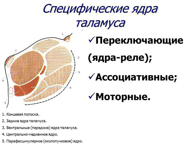
- Specific - switching afferent impulses to strictly localized areas of the cortex.
1.1. Relay (switching)
1.1.1.Touch(ventral posterior, ventral intermediate nucleus) switching of afferent impulses to sensory areas of the cortex.
1.1.2.Non-sensory - switching non-sensory information to the cortex.
- limbic nuclei(anterior nuclei) - subcortical center of smell. Anterior nuclei of the thalamus limbic cortex-hippocampus-hypothalamus-mamillary bodies of the hypothalamus - the anterior nuclei of the thalamus (Peypets reverb circle - the formation of emotions).
- Motor cores: (ventral) switch impulses from the basal ganglia, the dentate nucleus of the cerebellum, the red nucleus to motor and premotor area(transmission of complex motor programs formed in the cerebellum and basal ganglia).
1.2. Associative (integrative function, receive information from other nuclei of the thalamus, send impulses to the associative areas of the KGM, there is feedback)
1.2.1. Pillow nuclei - impulses from the geniculate bodies and non-specific nuclei of the thalamus, to the temporal-parietal-occipital zones of the CGM, involved in gnostic, speech and visual reactions (integration of the word with the visual image), perception of the body scheme. Electrical stimulation of the pillow leads to a violation of the naming of objects, the destruction of the pillow - a violation of the body scheme, eliminates severe pain.
1.2.2. Mediodorsal nucleus - from the hypothalamus, amygdala, hippocampus, thalamic nuclei, central gray matter of the trunk, to the associative frontal and limbic cortex. Formation of emotions and behavioral motor activity, participation in memory mechanisms. Destruction - eliminates fear, anxiety, tension, suffering from pain, but decreases initiative, indifference, hypokinesia.
1.2.3. Lateral nuclei - from the geniculate bodies, the ventral nucleus of the thalamus, to the parietal cortex (gnosis, praxis, body scheme.)
- Nonspecific nuclei - (intralaminar nuclei, reticular nucleus) signaling in all sections of KGM. Numerous incoming and outgoing fibers, an analogue of the RF stem - an integrating role between the brainstem, cerebellum and basal ganglia, neonatal and limbic cortex. Modulating influence, provide fine regulation of behavior, "smooth tuning" of GNI.
Metathalamus The medial geniculate bodies together with the inferior tubercles of the quadrigemina of the midbrain form the subcortical center of hearing. They play the role of switching centers for nerve impulses sent to the cerebral cortex. On the neurons of the nucleus of the medial geniculate body, the fibers of the lateral loop end. The lateral geniculate bodies, together with the superior tubercles of the quadrigemina and the pillow of the thalamus, are the subcortical centers of vision. They are communication centers at which the visual tract ends, and in which the paths that conduct nerve impulses to the visual centers of the cerebral cortex are interrupted.
Epithalamus The pineal gland is associated with the parietal organ of some higher fish and reptiles. In cyclostomes, it retained to a certain extent the structure of the eye; in anurans, it is found in a reduced form under the scalp. In mammals and humans, the pineal gland has a glandular structure and is an endocrine gland (hormone - melatonin).
The epiphysis (pineal gland) refers to the glands of internal secretion. It produces serotonin, from which melatonin is then formed. The latter is an antagonist of pituitary melanocyte-stimulating hormone, as well as sex hormones. The activity of the pineal gland depends on the illumination, i.e. the circadian rhythm is manifested, and this regulates the reproductive function of the body.
Hypothalamus
The hypothalamic region contains forty-two pairs of nuclei, which are divided into four groups: anterior, intermediate, posterior, and dorsolateral.
The hypothalamus is the ventral part of the diencephalon, anatomically consists of the preoptic region, the region of the optic chiasm, the gray tubercle and infundibulum, and the mastoid bodies. The following groups of nuclei are distinguished:
- Anterior group of nuclei (anterior to the gray nucleus) - preoptic nuclei, suprachiasmatic, supraoptic, paraventricular
- Intermediate (tuberal) group (in the region of the gray tubercle and infundibulum) - dorsomedial, ventromedial, arcuate (infundibular), dorsal hypotuberous, posterior PVN and own nuclei of the tubercle and infundibulum. The first two groups of nuclei are neurosecretory.
- Posterior - the nuclei of the papillary bodies (subcortical center of smell)
- Subthalamic nucleus of Louis (integration function
The hypothalamus has the most powerful network of capillaries in the brain and the highest level of local blood flow (up to 2900 capillaries per mm square). Capillary permeability is high, because The hypothalamus has cells that are selectively sensitive to changes in blood parameters: changes in pH, the content of potassium and sodium ions, oxygen tension, carbon dioxide. The supraoptic nucleus has osmoreceptors, the ventromedial nucleus has chemoreceptors glucose-sensitive in the anterior hypothalamus sex hormone receptors. Eat thermoreceptors. Sensory neurons the hypothalamus do not adapt, and are excited until one or another constant in the body is normalized. The hypothalamus carries out efferent influences with the help of the sympathetic and parasympathetic nervous systems, and endocrine glands. Here are the centers of regulation various kinds exchanges: protein, carbohydrate, fat, mineral, water, as well as centers of hunger, thirst, satiety, pleasure. The hypothalamic region is referred to the higher subcortical centers of autonomic regulation. Together with the pituitary gland, it forms the hypothalamic-pituitary system, through which the nervous and hormonal regulation is interfaced in the body.
In the hypothalamic region, endorphins and enkephalins are synthesized, which are part of the natural pain system and affect the human psyche.
Nerve pathways to the hypothalamus come from the limbic system, CGM, basal ganglia, RF trunk. From the hypothalamus to the Russian Federation, the motor and autonomic centers of the trunk to the autonomic centers of the spinal cord, from the mammillary bodies to the anterior nuclei of the thalamus, then to the limbic system, from the SOYA and PVN to the neurohypophysis, from the ventromedial and infundibular to the adenohypophysis, there are also connections with the frontal cortex and striped body.
Hormones SOYA and PVN:
- ADH (vasopressin)
- Oxytocin
Hormones of the mediobasal hypothalamus: ventromedial and infundibular nuclei:
Liberins (releasing) corticoliberin, thyroliberin, luliberin, folliberin, somatoliberin, prolactoliberin, melanoliberin
Statins (inhibins) somatostatin, prolactostatin and melanostatin
Functions:
- Maintenance of homeostasis
- Integrative Center for Autonomic Functions
- High Endocrine Center
- Regulation of heat balance (front nuclei - the center of heat transfer, rear - the center of heat generation)
- Regulator of the sleep-wake cycle and other biorhythms
- Role in eating behavior middle group nuclei: the lateral nucleus is the center of hunger and the ventromedial nucleus is the center of saturation)
- Role in sexual, aggressive-defensive behavior. Irritation of the anterior nuclei stimulates sexual behavior, irritation of the posterior nuclei inhibits sexual development.
- Center for the regulation of various types of metabolism: protein, carbohydrate, fat, mineral, water.
- It is an element of the antinociceptive system (pleasure center)